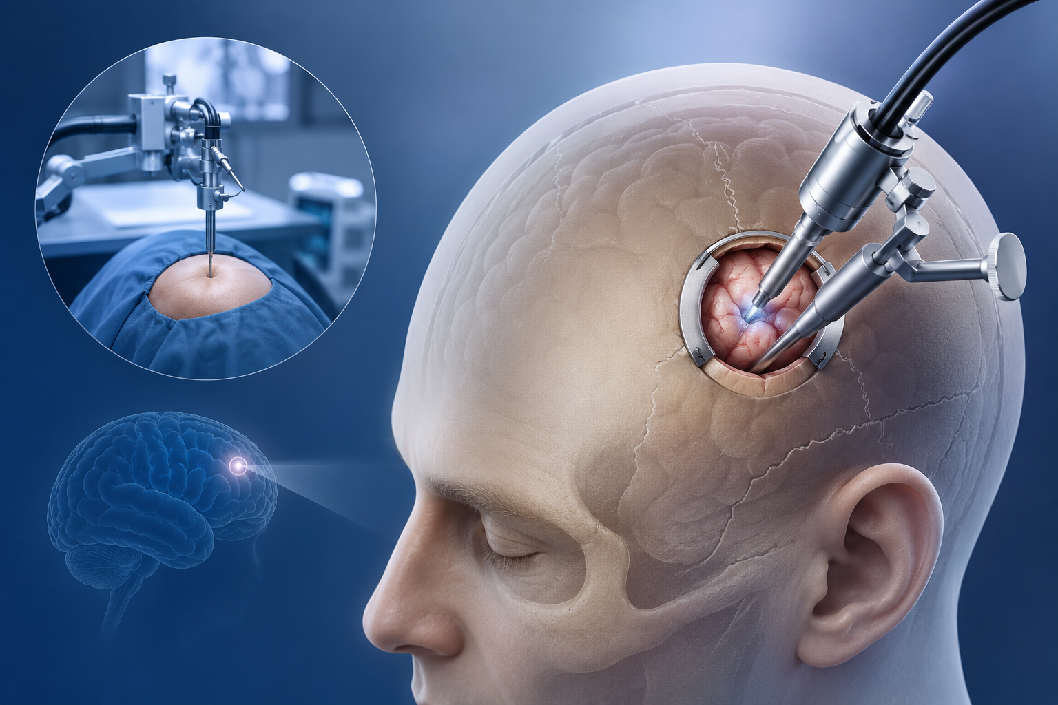
For decades, the mention of "brain surgery" evoked terrifying images: complete scalp shaving, large horseshoe incisions, removal of large skull bone flaps, and weeks of excruciating recovery in the ICU. Today, advances in 3D intraoperative neuro-navigation, high-definition endoscopes, and microsurgical instruments have ushered in a new era: Keyhole Brain Surgery.
What is Keyhole Brain Surgery?
Keyhole brain surgery (also known as Supraorbital Eyebrow Craniotomy or Minimally Invasive Craniotomy) is a surgical technique where tumors, aneurysms, and vascular lesions located in the anterior skull base and frontal lobes are removed through an incision measuring just 3 to 4 centimeters hidden within the natural contour of the patient's eyebrow.
Rather than making a large opening, the neurosurgeon utilizes the geometric principle of a keyhole: while the surface aperture is small, the visual cone inside expands significantly as high-magnification surgical operating microscopes and angled 4K endoscopes illuminate deep brain structures without compressing healthy brain tissue.
Keyhole vs. Traditional Open Craniotomy: At a Glance
| Parameter | Traditional Open Craniotomy | Keyhole Eyebrow Craniotomy |
|---|---|---|
| Hair Shaving | Extensive or Full Head Shave | Zero Head Shaving (Eyebrow Hair Preserved) |
| Incision Size | 15 cm to 25 cm curved scalp incision | 3 cm to 4 cm hidden in eyebrow crease |
| Skull Bone Opening | Large flap (8 to 12 cm) | Small keyhole window (approx. 2 cm) |
| Brain Retraction | Higher retraction of normal frontal lobe | Minimal to zero retraction via natural corridors |
| ICU & Hospital Stay | 2-3 days ICU, 7-10 days in hospital | Overnight ICU, discharged in 48-72 hours |
| Cosmetic Outcome | Visible scalp scar, possible indentation | Virtually invisible scar once eyebrow hairs heal |
Which Conditions Can Be Treated via Keyhole Brain Surgery?
Dr. Agnetia Vinoth regularly evaluates patients across Chennai, Tamil Nadu, and international referrals for keyhole approaches. Ideal conditions include:
- Anterior Skull Base Meningiomas: Olfactory groove, planum sphenoidale, tuberculum sellae, and sphenoid wing meningiomas.
- Craniopharyngiomas: Suprasellar tumors compressing the optic chiasm.
- Metastatic Brain Tumors: Lesions located in the frontal lobes or skull base.
- Cerebral Aneurysms: Selected anterior communicating artery (ACom) and internal carotid artery (ICA) aneurysms amenable to microsurgical clipping.
- Epidermoid and Dermoid Cysts: Located in the basal cisterns.
The Role of 3D Neuro-Navigation ("Brain GPS")
Operating through a 2 cm bone window requires sub-millimeter precision. Dr. Agnetia Vinoth utilizes optical 3D neuro-navigation. Prior to surgery, high-resolution volumetric MRI and CT angiography scans are loaded into the navigation console. During surgery, infrared cameras track surgical instruments relative to the patient's anatomy in real time with 0.5 mm spatial accuracy.
This guarantees that the exact shortest trajectory to the tumor is taken, critical optic nerves and major cerebral arteries are protected, and complete tumor resection is verified before wound closure.
Have You Been Diagnosed with a Brain Tumor?
Before undergoing extensive craniotomy with full head shaving, get an expert surgical second opinion from Dr. Agnetia Vinoth to discover if your lesion is eligible for a minimally invasive eyebrow keyhole approach.
Frequently Asked Questions (FAQ)
Will my eyebrow hairs be shaved or permanently damaged?
No. The eyebrow hair is never shaved. The skin incision is placed parallel to the hair follicles within the brow margin. Once healed, eyebrow regrowth completely conceals the fine surgical scar.
Is keyhole brain surgery as safe as traditional surgery?
When performed by a specialized skull base neurosurgeon, keyhole surgery has equivalent or superior tumor clearance rates with significantly lower complication rates, lower blood loss, zero muscle atrophy, and lower risk of surgical site infections.
How fast can I resume normal life and work?
Most patients walk comfortably the day after surgery and are discharged within 48 to 72 hours. Desk work and routine light daily activities can typically be resumed within 2 to 3 weeks, compared to 8 to 12 weeks for traditional craniotomies.