The skull base is the delicate anatomical floor upon which the brain rests, separating the brain from the nasal cavity, ear canals, and facial skeleton. Deep within this complex crossroads sits the pituitary gland—the body's master endocrine organ—surrounded by the optic nerves (responsible for eyesight) and the carotid arteries.
When a tumor develops in this region, such as a pituitary macroadenoma, craniopharyngioma, or chordoma, patients often experience progressive vision loss (tunnel vision or bitemporal hemianopsia), debilitating headaches, and severe hormonal imbalances. Historically, reaching these tumors required opening the skull. Today, with the Expanded Endoscopic Endonasal Approach (EEA), surgery is performed completely through the nostrils without a single external cut.
How Does Endoscopic Endonasal Surgery Work?
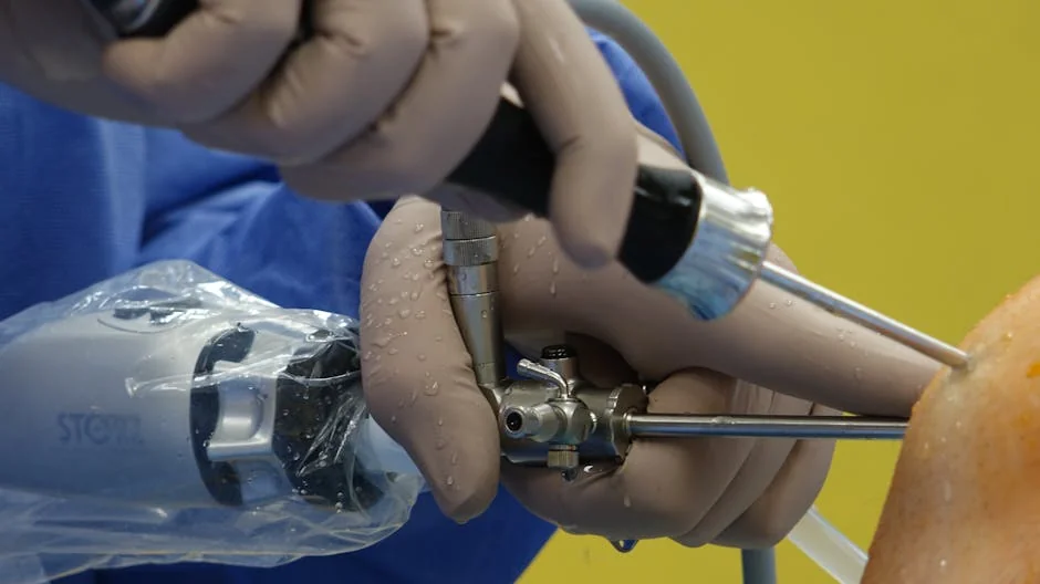
Using ultra-thin rigid 4K endoscopes (measuring only 4 mm in diameter), the surgical team navigates through the natural nasal corridors into the sphenoid sinus. A small window is created in the thin bone beneath the pituitary fossa (sella turcica).
The angled endoscope brings light and magnification directly up against the tumor, giving the neurosurgeon a panoramic, close-up view of the junction between tumor tissue, the healthy normal pituitary gland, and the stretched optic chiasm. Specialized microsurgical curettes and ultrasonic aspirators are used to gently decompress the optic nerves and remove the tumor.
Key Advantages of the Endoscopic Endonasal Approach
Zero External Incision
No skin cuts, no scars, no facial distortion, and zero head shaving. The entire procedure is performed entirely via the nostril cavities.
Immediate Vision Decompression
Because the optic chiasm is directly decompressed from below without needing to lift or push the brain, many patients report noticeable improvement in visual acuity within 24 to 48 hours post-op.
Maximum Pituitary Gland Preservation
High-definition magnification allows the surgeon to clearly identify and preserve normal compressed gland tissue, reducing long-term dependence on hormone replacement therapies.
Rapid 3 to 4 Day Recovery
Without brain retraction or craniotomy pain, patients ambulate the following day and are discharged home within 72 to 96 hours with minimal discomfort.
What About Acoustic Neuromas (Vestibular Schwannomas)?
Acoustic neuromas are non-cancerous skull base tumors that originate on the vestibular nerve in the cerebellopontine angle (CPA), adjacent to the facial nerve (cranial nerve VII) and inner ear hearing apparatus.
For acoustic neuromas, Dr. Agnetia Vinoth employs a Retrosigmoid Keyhole Approach with continuous intraoperative Facial Nerve EMG Monitoring. During every millimeter of microsurgical dissection, auditory and facial nerve signals are monitored in real time, dramatically maximizing the likelihood of preserving facial muscle symmetry and hearing function.
Specialist Evaluation for Pituitary or Skull Base Tumors
Facing vision decline, prolactin elevation, or an incidental skull base MRI finding? Consult Dr. Agnetia Vinoth for an individualized multidisciplinary review in Chennai or via international video consultation.
Frequently Asked Questions (FAQ)
How do surgeons prevent brain fluid (CSF) leaks after surgery?
Multilayer skull base reconstruction is performed. Using vascularized nasoseptal flaps, bio-absorbable dural matrices, and tissue glues, a watertight biological seal is reconstructed, reducing CSF leak risks to under 2%.
Will my sense of smell or breathing be affected?
Temporary nasal congestion and mild scabbing are normal for 2 to 3 weeks while mucous membranes heal. Saline nasal rinses are prescribed. Long-term olfactory function and nasal breathing are typically completely preserved.
Can non-functioning pituitary tumors recur?
When gross total resection is achieved, recurrence rates are exceptionally low. Routine annual surveillance MRIs and hormonal blood panels are conducted to ensure lifelong disease control.